Canada’s First Electron Microscope 1938
Professor Burton and grad students Hillier and Prebus developed the first practical electron microscope that focused a beam of electrons for illumination
As with most inventions in the world, advanced ideas build on the information of other bright ideas, and innovation climbs up to stand on the shoulders of previous innovation. Such was the case with the practical electron microscope built in 1938 at the University of Toronto.
Three years earlier, Professor E.F. Burton, Chairman of the Physics Department, was at a meeting in Berlin, Germany to discuss the useful possibilities of electron microscopy. Observed at the originator of the instrument, German scientist Ernst Ruska built an electron microscope in 1931. He had trouble with refining his design, resulting in specimens destroyed by the hot beam of the microscope. “After attending the meeting Burton became certain that, once perfected, the electron microscope would be a key tool in biological and medical research,” said Julie Stoehr in The Undiscovered Country: Development of the Electron Microscope (Quasar, University of Alberta). Burton “returned to Canada determined to construct such an instrument.”
Graduate Students Begin Electron Microscope
Inspiring the interest of graduate students over several years, Burton enlisted Cecil Hall, Albert Prebus and James Hillier to construct a microscope. It was to be a radical change from the standard light microscope. Their task was “to build a compound, transmission, magnetic electron microscope and to apply it to biological and colloidal materials,” according to The Electron Microscope: A Personal Recollection, by John H.L. Watson of the University of Toronto. As their foundation, the three young men used the thesis Hall wrote on designing a rudimentary electron microscope in 1935.
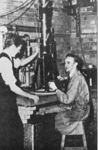
Hillier and Prebus working with the 1938 Electron Microscope
Work began in January 1938 with two shifts, day and evening, to create the components of the equipment. Prebus and Hillier “decided on a bold course of action,” said John H. Reisner in “Advances in Electrons and Electronic Physics” (Academic Press Inc., San Diego 1973). “They elected to build their transmission microscope in a single jump, avoiding a slower and much more cautious step-by-step building and testing of its several components.” Their successful risk saved a year or more of development time.
The Electron Microscope Enhanced Specimen Magnification
Testing and fine-tuning the electron microscope, the team was able to reach a primary magnification of 30,000 times, without destroying the examined specimen. (Light microscopes in the mid-1900s reached 2,000 times magnification.) Burton was able to publish reports on the new electron microscope in popular magazines and in scientific journals only a year after the project began.
The first electron microscope in 1938 was large, standing at 6 feet tall on a base. Built in “six sections, each joined to the other by vacuum-tight (it was hoped!) plane-lapped, vacuum-greased seals, as an upright vertical column of the six sections,” noted Watson. “This arrangement allowed horizontal, sliding adjustment of each section for alignment of optical axes and for selection of specimen areas.” As dramatically oversimplified description, the top section held the electron gun and condenser, the second section held the specimen chamber, sitting upon the objective electromagnetic lens of the third section.
Unshielded Power a Hair-Raising Experience
The fourth microscope section housed a fluorescent screen for viewing the image projected by the magnifying lens in the fifth section. (The fourth section was shielded in a brass tube to protect the beam from external magnetic fields and electrical interference.) The lens was powered by 24-volt storage batteries with rheostat adjusters. Under the control of an operator, the camera in the sixth section captured micrographs of the magnified specimen. (A micrograph is a photo taken by a microscope.) The microscope itself was supplied by unprotected high voltage electricity. Once, while adjusting the electron microscope, Watson noted, the “unshielded 45 KV voltage [was] so close that the hairs on the back of my hands, or my youthful head were raised,” said Watson. The early electron microscope had many scientific and medical applications, including examination of bacteria and viruses, the structure of leaves and plants, and for the study of physics.
The ground-breaking men responsible for constructing the University of Toronto’s electron microscope were recruited to work at large American companies. Hall left Canada in 1937, just as the university project was getting underway, to build an electron microscope for Eastman Kodak Company. The Radio Corporation of America (RCA) hired Hillier on completion of his PhD in 1941; he constructed the company’s first commercial electron microscope. Joining Ohio State University as a faculty member, Prebus built an electron microscope for physics research at the institution.
Microscope on Permanent Museum Display
In 1988 on the 50th anniversary of its invention, the first practical electron microscope was commemorated by Canada Post with a 37-cent postage stamp, part of a series entitled “Canadian Innovations in Science and Technology.”

Canada Post Stamp, Issued July 1988: The First Electron Microscope 1938
The microscope is now on permanent display at Canada Science and Technology Museum in Ottawa, Ontario. The fascinating device constructed of glass tubes, metal, wire and string was proudly unveiled on April 7, 2011 by David Pantalony, the Museum’s Curator of Physical Sciences and Medicine. Electron microscope development continues to advance, building on the inventions of the past and reaching for the innovations of the future.
Sources
- Reisner, John H., essay, “An Early History of the Electron Microscope,” Advances in Electrons and Electronic Physics, edited by Peter W. Hawkes, Academic Press Inc., San Diego 1973.
- Stoehr, Julie, The Undiscovered Country: Development of the Electron Microscope, Quasar, University of Alberta Accessed April 7, 2011
- Watson, John H.L., The Electron Microscope: A Personal Recollection, Physics Department, University of Toronto Accessed April 7, 2011
This article first appeared on Suite101.com on April 9, 2011. Copyright Susanna McLeod